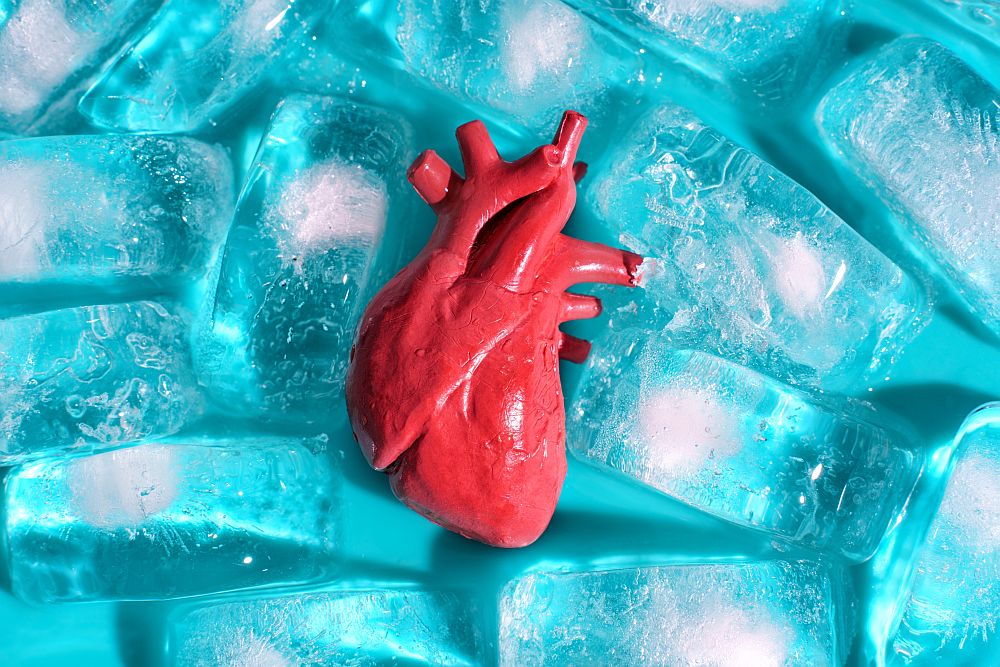
[Image above] Successful root canal therapy requires all the materials used in the treatment to interact well with the complex tissue environment within and surrounding the tooth. A new study investigated the biocompatibility of two newer bioceramic sealers. Credit: Gustavo Fring, Pexels
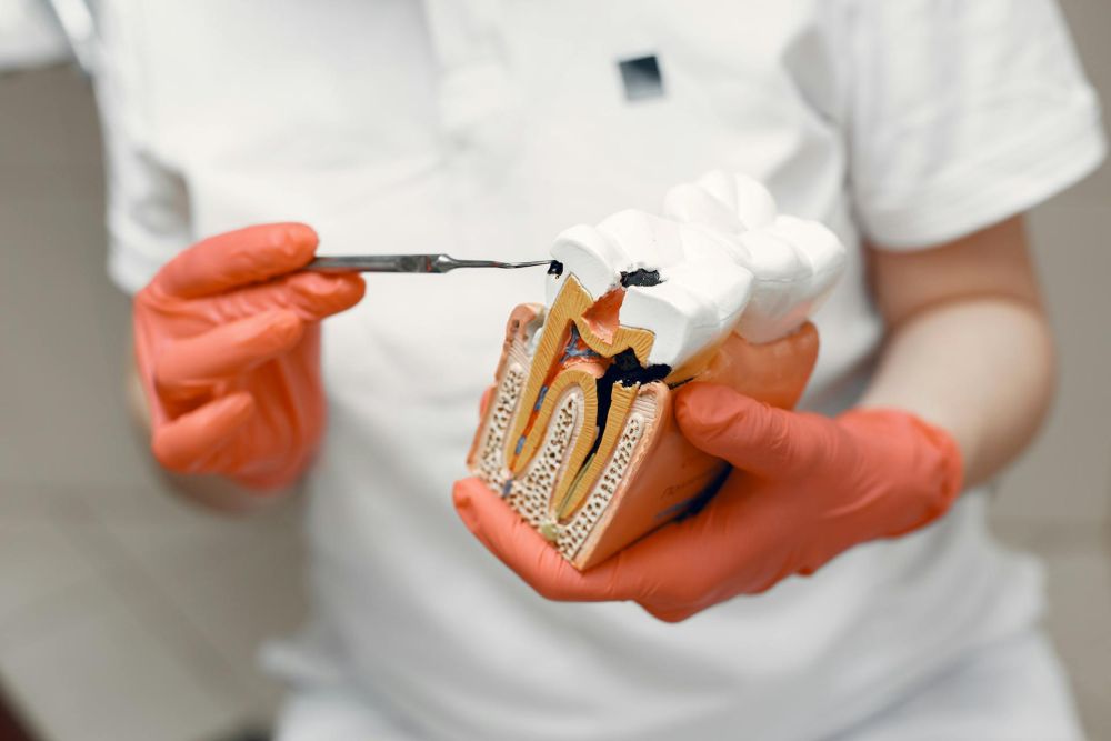
Though many people still view root canals as one of the most painful dental procedures, advancements in medical technology have made this treatment, which is used to remove inflamed or infected pulp (soft tissue) on the inside of a tooth, typically no more painful than getting a filling.
As shown in the video below, the multistep root canal procedure begins with the dentist using a small, rotating file to extract the damaged pulp from the tooth’s root canal. A filling material, typically the natural rubber gutta-percha, is then placed inside the empty canal, and a sealing material is injected to fill the remaining space.

Credit: rootcanalspecialists, YouTube
Currently, a hydrophobic epoxide-amine resin sealer called AH Plus, produced by U.S. dental equipment and consumables manufacturer Dentsply Sirona, is considered the gold standard sealing material for root canal treatments due to its long-term sealing integrity, low shrinkage, and high radiopacity (ability to be seen in X-ray images). But it does face some disadvantages, such as difficulties in connecting to the gutta-percha and questionable cementation to the tooth canal wall in the presence of moisture.
Among other sealer types, bioceramic sealers entered the scene during the 1990s, but the improved flowability of recent generations brought this sealer type back into the spotlight. In contrast to the standard AH Plus formulation, bioceramic sealers typically bond better with the gutta-percha and tooth canal wall due to bioactivity. They also expand slightly rather than shrink upon setting, improving the final seal as well.
Despite the well-known sealing properties, surprisingly little is known about the interaction of some newer bioceramic sealers with the periapical tissues (i.e., the tissues surrounding the tip of a tooth root). While ideally all the sealer would stay inside the tooth canal, various factors can lead it to escape into the periapical tissues. So, while the interaction of sealer and periapical tissues is not a priority during initial development, it is an important consideration as bioceramic products enter commercial practice.
In a recent open-access paper, researchers from several universities in Egypt investigated the biocompatibility of two newer bioceramic sealers on the market: CeraSeal and NeoSEALER Flo.
CeraSeal is produced by Korean medical device manufacturer Meta-Biomed, while NeoSEALER Flow is produced by U.S. bioactive dental materials manufacturer Avalon Biomed. Like many root canal sealers, these sealers contain calcium silicate as a bioactive component, which is antimicrobial due to its high pH. Regarding radiopacifiers, or the materials that ensure visibility during radiographic assessment of root canal fillings, CeraSeal relies mainly on zirconium dioxide (45–50%) and NeoSEALER Flo relies mainly on tantalite (50%).
The researchers investigated the interaction of these sealers with human gingival fibroblasts (HGFs), the primary cell type in gingival tissue (the gums). These cells play a crucial role in maintaining tissue integrity and wound healing, as well as in modulating the immune response to oral pathogens.
They cultured and treated HGFs with solutions of the two sealers at different dilutions: 25%, 50%, 75%, and 100%. The treated cells then were left in an incubator for up to 7 days, and cytotoxic effects and inflammatory response were determined using MTT assay and qRT-PCR, respectively. Samples with AH Plus were also created and tested to serve as a control.
The MTT assay results revealed that both CeraSeal and NeoSEALER Flo bioceramic sealers exhibited concentration-dependent cytotoxicity on HGFs. At the highest concentration, CeraSeal showed a 35% reduction in cell viability compared to the control, whereas NeoSEALER Flo showed a 45% reduction.
Furthermore, the qRT-PCR indicated that exposure to CeraSeal and NeoSEALER Flo resulted in elevated levels of pro-inflammatory cytokine production in HGFs, with NeoSEALER Flo causing a more pronounced inflammatory response. While some inflammation can be beneficial for healing, too much inflammation is detrimental.
Based on these findings, the researchers report a slight preference for CeraSeal biocompatibility. They attribute this result to CeraSeal’s refined formulation, which “may minimize the release of harmful substances,” and its improved setting properties, which “may reduce the solubility and leaching of cytotoxic components.”
However, they emphasize that because the study was conducted on Teflon disks, as well as only including the HGF cell type, it does not reflect the complex environment of in vivo periodontal tissues and so cannot serve as an indicator of clinical success. As such, future studies should use in vivo animal models “to assess the biological responses in more physiologic relevant context,” they write.
The open-access paper, published in BMC Oral Health, is “Comparative analysis of the inflammatory response of human gingival fibroblasts to NeoSEALER Flo and CeraSeal bioceramic sealers: an in vitro study” (DOI: 10.1186/s12903-025-05692-1).
Update 05/13/2025 – Slight updates to the text to more accurately reflect the conclusions of the study.