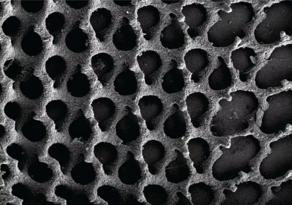
[Image above] Samples of silicon-substituted hydroxyapatite, a synthetic bone material, were exposed to gamma radiation to investigate the effects on its electrical properties. Credit: Machuga et al., International Journal of Ceramic Engineering & Science
Exposure to high-energy radiation, such as X-rays and gamma rays, can lead to exceedingly negative health effects when it occurs in high doses. But fortunately, on Earth, the overall levels of terrestrial external radiation are low. As such, humans typically only encounter high-energy radiation in the context of controlled medical facilities, which allows for exposure to be easily moderated.
Outer space, however, is a different matter. The space environment is filled with fast-moving ions with sufficient energy to change or break DNA molecules. The Earth’s atmosphere and magnetic field prevent this space radiation from reaching the surface. But as humans venture deeper into space away from the Earth’s protective barrier, exposure to high-energy radiation can no longer be easily moderated.
Understanding the long-term effects of this exposure is necessary to realizing the dream of space travel and colonization. In a recent open-access article, researchers at the University of Maryland, Baltimore County (UMBC) worked with colleagues at NASA to investigate the effects of high-energy radiation on hydroxyapatite, an important bone material.
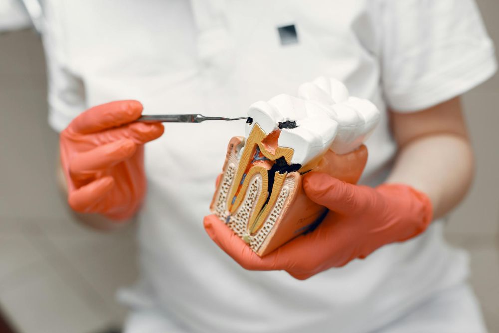
Hydroxyapatite is a naturally occurring calcium phosphate mineral found in bones and teeth. Since the 1950s, doctors have used hydroxyapatite derived through both natural and synthetic sources for bone regeneration treatment.
For hydroxyapatite to adhere well with natural bone, its electrical properties are an important factor. It is known that the electrical properties of materials can change upon exposure to radiation, so the UMBC researchers investigated how hydroxyapatite reacts to such exposure.
For this study, the researchers synthesized and analyzed silicon-substituted hydroxyapatite, which features silicon atoms in place of phosphorus atoms within the apatite structure. Compared to natural hydroxyapatite, the incorporation of silicon results in materials with comparable biocompatibility and mechanical properties but with improved bioactive behavior, as demonstrated by both in vitro and in vivo tests.
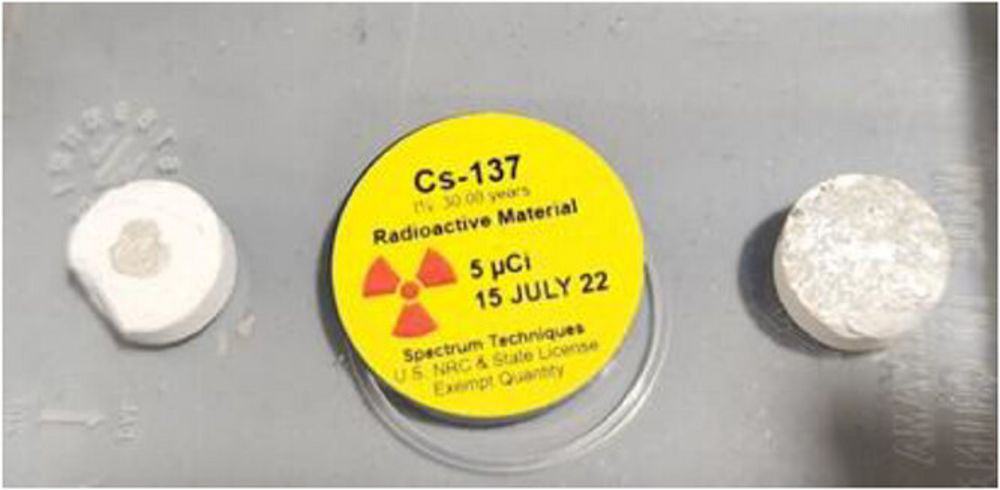
They exposed the silicon-substituted hydroxyapatite sample to gamma radiation for up to 100 hours using Cs-137 as the radiation source. Cs-137 only has a radiation dose of 5 µm curie, but it “was [the] only allowable dose for our laboratories,” they explain.
Even though they could not use a higher radiation dose, the researchers observed significant changes in the sample’s dielectric constant and resistivity, with the former greatly increasing and the latter greatly decreasing. Maximum change occurred within the first 20 hours of exposure, suggesting that longer exposure times may not be necessary in future studies.
The researchers note that a material’s dielectric constant and resistivity depend on the amount of oxidation and grain boundaries in the sample. As such, modifying the processing temperature and time for the sample, as well as adding small amounts of other materials to the mix, may help counteract the resulting changes that occur during high-energy irradiation.
The open-access paper, published in International Journal of Ceramic Engineering & Science, is “Effect of high-energy radiation on electrical properties of synthetic bone materials” (DOI: 10.1002/ces2.10202).