Ptychography – High resolution microscopy using mathematics instead of lenses
Ptychography is a new approach to microscopy that eschews lenses. Instead, diffraction patterns are collected and reconstructed using algorithms that work backwards to create the image.
When we were learning about Fourier transformations in my college linear algebra class, the instructor wrote a big matrix on the board, pointed to it and said, “That’s a lens.” I remember being surprised at seeing something I’d always thought of as having to be tangible expressed as a mathematical abstraction.
What if you could go the other way? That is, what if mathematics were applied to diffracted electron beams and reconstructed into images instead of applying lenses to electron beams on their way to the specimen?
That is the approach taken by a group led by John Rodenburg from the University of Sheffield (U.K.). They may be on the verge of revolutionizing the field of high-resolution microscopy by, basically, using mathematics to construct images from diffraction patterns, eliminating the need for optics and their limitations. In a recent paper in Nature Communications, the authors explain, “Here we demonstrate a form of diffractive imaging that unshackles the image formation process from the constraints of electron optics, improving resolution over that of the lens used by a factor of five …”
They say the method, called electron ptychography, “has no fundamental experimental boundaries: further development of this proof-of-principle could revolutionize sub-atomic scale transmission imaging.” The best resolution possible in a fully optimized SEM set-up would be about 1.2 nanometers, they say in the paper. Resolutions of about 0.24 nanometers (atomic scale) are achievable with ptychography.
In a press release from the university, Rodenburg explains how the image is constructed.
We measure diffraction patterns rather than images. What we record is equivalent to the strength of the electron, X-ray or light waves which have been scattered by the object – this is called their intensity. However, to make an image, we need to know when the peaks and troughs of the waves arrive at the detector – this is called their phase.
The key breakthrough has been to develop a way to calculate the phase of the waves from their intensity alone. Once we have this, we can work out backwards what the waves were scattered from: that is, we can form an aberration-free image of the object, which is much better than can be achieved with a normal lens.
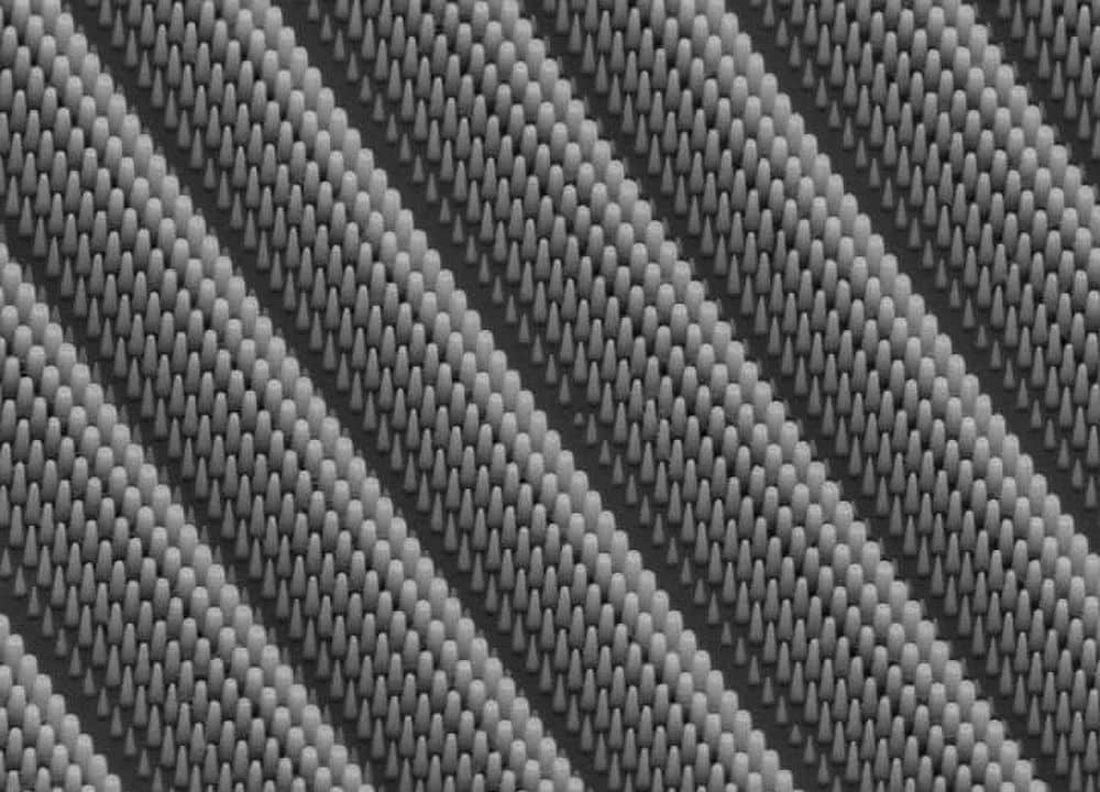
Although the paper describes the method for electron microscopy, ptychography can be applied to microscopy using other illumination sources including visible light, producing images such as those above. This would be advantageous in the biological sciences. Without lenses closing in on them, cells can be observed without disruption, for example.
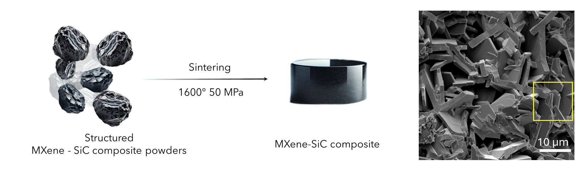
The method is still in its proof-of-concept stage. The paper presents images of gold particles to demonstrate the capability of the method, but it is expected to be applicable to ceramic materials, too. In an email, Rodenburg said the group had not looked at any ceramic specimens yet, “However, it is certainly true to say that electron imaging is key for the study of ceramics, so certainly as we (and, hopefully, others) adopt and improve the approach it will have significant impact in the study of ceramics.”