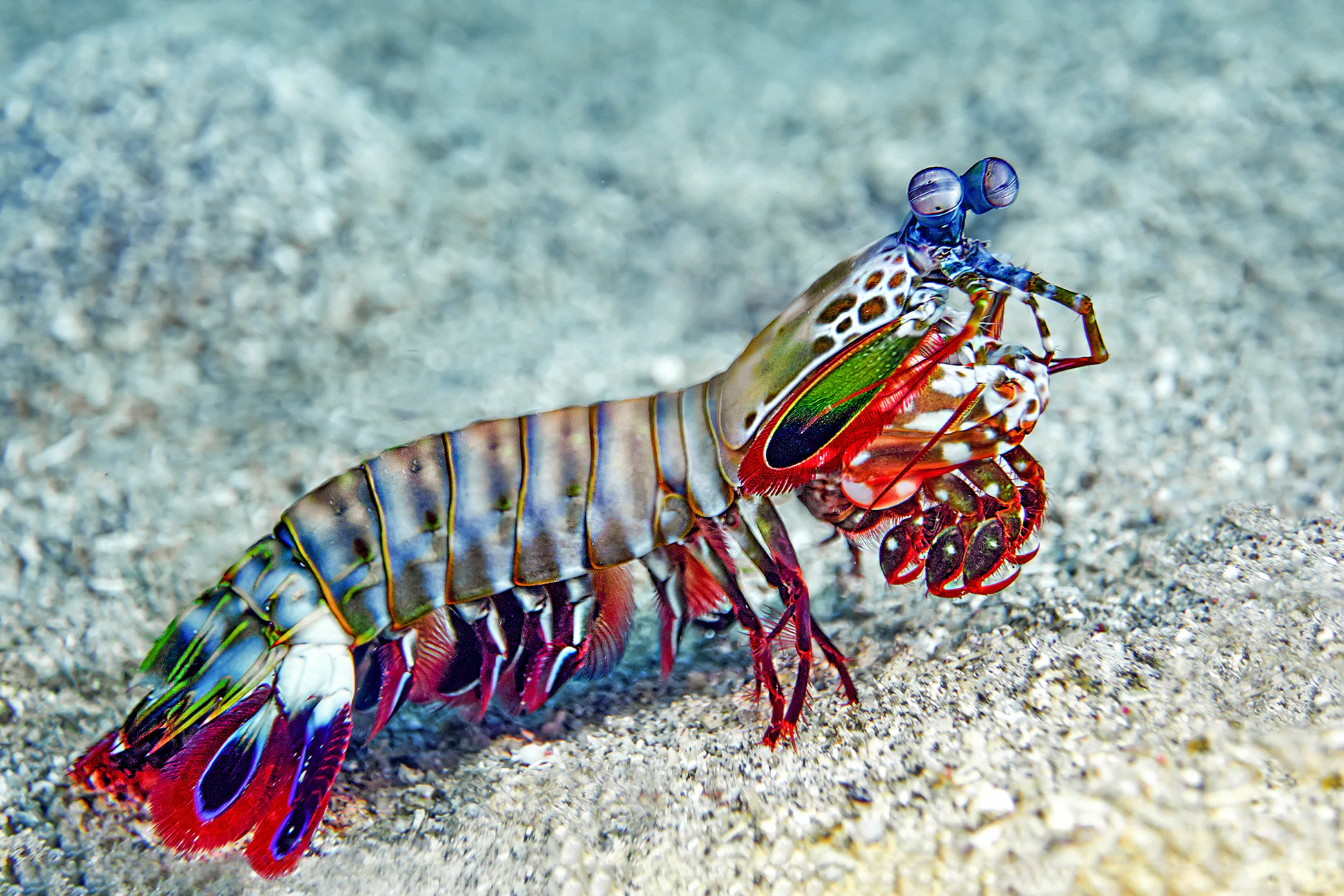
[Image above] Mantis shrimp not only see in technicolor, they dress in technicolor as well. Credit: Dorothea OLDANI, Unsplash
Nature is inspiring.
Take, for example, Mariah Reading’s paintings of the beautiful national park landscapes surrounding discarded pieces of trash, which serve as the canvas for her works. Or the work of artistic scientists like Balaram Khamari, who doesn’t have to stray far from the microbiology bench to paint masterpieces, using microbes as his medium and agar plates as the canvas.
Of course, artists aren’t the only ones—scientists also often turn to nature for inspiration when designing structures, materials, and more. One bizarre little creature in particular seems to be serving as the muse for a handful of scientific innovations recently: the mantis shrimp.
While neither a mantis nor a shrimp, these carnivorous marine crustaceans have some impressive evolutionary adaptations. For starters, mantis shrimp have some of the most complex eyes found in nature, equipped with more than a dozen kinds of different photoreceptors (we humans have just three) and able to detect not only visible and UV light but also polarized light as well. In other words, mantis shrimp see the world in technicolor.
Beyond their incredible eyes, mantis shrimp are perhaps even more famous for their powerful punch. Also known as the “thumb splitter” and notorious for breaking the glass of aquarium walls, the mantis shrimp punches above its weight—it can propel its appendages forward and away from its body at an explosive 50 mph, delivering blows with such intense force (upwards of 1,500 N) that the mantis shrimp can smash apart crab exoskeletons and mollusk shells.
In addition to the impressive capacity to generate such explosive force, this punching ability of the mantis shrimp also means that the creature’s fists—called dactyls—are equipped with some seriously tough materials to withstand such force. And that makes them perfect inspiration for engineers looking to design fracture-resistant materials.
Building Bouligand with bacteria
Like many biomaterials, the mantis shrimp’s dactyls garner strength from a structure that efficiently dissipates energy and resists fracturing. Previous studies showed this strength stems from a combination of different structural adaptations, including a nanoparticle structure that dissipates energy and a herringbone pattern in the outer layer that prevents cracking.
Inside the dactyls, layers of materials are arranged in an offset helical pattern called a Bouligand shape—twisted like a fancy stack of cocktail napkins—that further helps dissipate energy and prevent fracture.
While scientists have long known about the strength of this Bouligand shape, its complexity makes it difficult to fabricate synthetically, especially in mineralized materials. Now, a team of researchers at the University of Southern California and University of California, Irvine devised a novel strategy to fabricate such structures in the lab, by harnessing nature herself to help grow these complex patterns.
“We have been amazed by the sophisticated microstructures of natural materials for centuries, especially after microscopes were invented to observe these tiny structures,” Qiming Wang, an engineer and researcher at the University of Southern California Viterbi School of Engineering and senior author of the new work, says in a USC press release. “Now we take an important step forward: We use living bacteria as a tool to directly grow amazing structures that cannot be made on our own.”

The living materials mimic a Bouligand structure found in nature, including in the mantis shrimp’s dactyls. Credit: Qiming Wang, USC
To create the mantis shrimp-inspired structures, the researchers first 3D printed a framework structure consisting of layers of open polymer lattices. Then, they seeded the structures with living bacteria, Sporosarcina pasteurii, which secrete the enzyme urease.
In the presence of calcium ions and urea, urease catalyzes formation of the mineral calcium carbonate, the same stuff that helps make bone, teeth, and seashells strong. (And some bricks as well!) So when the researchers dunked the bacteria-laden polymer lattices into a solution containing urea and calcium ions, the bacteria’s urease catalyzed mineralization of calcium carbonate within the framework, creating an increasingly solid structure.
By stacking similar layers of the framework in an offset helical pattern, the researchers showed that they could catalyze creation of a mineralized calcium carbonate Bouligand structure—simply by giving the bacteria a place to grow and some food to eat.

Credit: Qiming Wang, YouTube
“The key innovation in our research is that we guide the bacteria to grow calcium carbonate minerals to achieve ordered microstructures which are similar to those in the natural mineralized composites,” Wang says in the release.
The structures revealed a promising combination of high strength, fracture resistance, and energy dissipation. Testing revealed the bacteria-built Bouligand structures provided strength by helping dissipate energy and by preventing cracks from propagating within the structure.

A) Schematic of the lattice structure. B) Growth of the calcium carbonate mineralized structure over time. C) Optical microscope images of the lattice mineralization. D) SEM images of the lattice mineralization. E) Mineral thickness growth on the lattice over time. F) Compressive stress v. strain of the mineralized lattice over time. G) Effective stiffness of the mineralized lattice over time. Credit: Qiming Wang, USC
Wang explains in a Wired article: “This kind of microstructure makes sure that this kind of composite is very tough…When you have a crack, that crack will propagate in the twist pattern to dissipate the energy inside the material.” The article continues: “In fact, the material absorbs more energy than natural nacre (mother of pearl), which gives some shells their strength, and also beats existing artificial materials, Wang and his colleagues say.”
In addition to representing a proof-of-concept for “3D‐architectured hybrid synthetic–living materials with living ordered microstructures and exceptional properties,” as the authors call them in the paper’s abstract, the team sees diverse potential applications, including lightweight body armor or vehicle armor. “This material could resist bullet penetration and dissipate energy from its release to avoid damage,” postdoc and co-author Yipin Su says in the release.
And because the materials are grown with the help of live bacteria, the researchers also see a potential direction toward self-healing materials. “An interesting vision is that these living materials still possess self-growing properties,” Wang says in the release. “When there is damage to these materials, we can introduce bacteria to grow the materials back. For example, if we use them in a bridge, we can repair damages when needed.”
The paper, published in Advanced Materials, is “Growing living composites with ordered microstructures and exceptional mechanical properties” (DOI: 10.1002/adma.202006946).
Seeing an optical advantage
It’s not just the mantis shrimp’s dactyls that are inspiring innovation, however—the creature’s highly complex eyes also helped another team of researchers see a new strategy for developing compact yet sophisticated optical sensors.
Scientists at North Carolina State University developed small optical sensors capable of advanced hyperspectral and polarimetric imaging—previously only achieved in much larger, more complex sensors—which could expand the use of compact devices such as smartphones for more precise analysis of scientific imaging.
“Lots of artificial intelligence (AI) programs can make use of data-rich hyperspectral and polarimetric images, but the equipment necessary for capturing those images is currently somewhat bulky,” Michael Kudenov, co-corresponding author of a paper describing the work and associate professor of electrical and computer engineering at NC State, says in a university news story. “Our work here makes smaller, more user friendly devices possible. And that would allow us to better bring those AI capabilities to bear in fields from astronomy to biomedicine.”

Stomatopod Inspired Multispectral and POLarization sensitive (SIMPOL) image showing spectral imaging of a scene containing objects with different colors, as well as the letters NCSU, which contain different polarization states. Image credit: Ali Altaqui, NC State
The organic electronic sensor, which the researchers named “SIMPOL” for Stomatopod Inspired Multispectral and POLarization sensitive, mimics the complex eye of the mantis shrimp, also called a stomatopod.
Like the mantis shrimp eye, the sensor can detect polarized light and also can detect visible light in narrower bands—10 times narrower (hence, hyper-spectral)—than other such sensors, allowing much more precise detection. And detection of polarized light with polarimetric imaging can provide important information about an object’s surface geometry.
While the technology isn’t going to help you take a better picture from your smartphone, it could open up significant possibilities for scientific imaging across diverse fields.
“Our work demonstrates that it is possible to create small, efficient sensors that can simultaneously capture hyperspectral and polarimetric images,” says Brendan O’Connor, co-corresponding author of the paper and an associate professor of mechanical and aerospace engineering at NC State. “I think this opens the door to a new breed of organic electronic sensing technologies.”
The open-access paper, published in Science Advances, is “Mantis shrimp–inspired organic photodetector for simultaneous hyperspectral and polarimetric imaging” (DOI: 10.1126/sciadv.abe3196).
Author
April Gocha
CTT Categories
- Material Innovations
- Nanomaterials
- Optics


