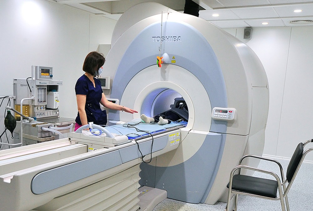
[Image above] An example of a magnetic resonance imaging scanner by Japanese multinational conglomerate Toshiba. Researchers in Russia recently proposed a modified scanner design that may examine small areas more effectively. Credit: Mos.ru, Wikimedia (CC BY 4.0)
When it comes to medical technologies, one of the most iconic devices is a magnetic resonance imaging (MRI) scanner.
MRI is a noninvasive medical imaging technique used to diagnose a variety of conditions, from torn ligaments to tumors. The technique relies on radio waves and a powerful magnet linked to a computer to create detailed pictures of areas inside the body. Compared to CT scans and X-rays, MRI does not use potentially harmful ionizing radiation.
A conventional MRI scanner, which is illustrated below, resembles a large tube with a table in the middle for a patient to lay on. Within the tube, radiofrequency (RF) waves are transmitted to the patient via a large, human-sized birdcage coil, which consists of two circular conductive loops (called end rings) connected by several conductive straight elements (called rungs or legs). The MRI signal is detected by multiple receive-only surface coils that are located directly on a patient, and a cabling system delivers those signals to a spectrometer.

An illustration of a conventional data acquisition scheme in clinical MRI scanners. Credit: Shchelokova et al., Nature Communications (CC BY 4.0)
The conventional MRI design provides acceptable image quality for examinations of the whole body as well as certain body parts and organs. But for examinations that focus on smaller areas such as the head, spine, joints, and breasts, “The RF magnetic field of the body birdcage coil is distributed over its total volume, requiring higher power during excitation that limits the coil transmit efficiency for small areas,” researchers write in a recent open-access paper.
The researchers come from several institutes and companies in Russia, and they are led by assistant professor Alexey Slobozhanyuk and chief researcher Anna Andreychenko from ITMO University in Saint Petersburg.
In their paper, the researchers note that use of dedicated transceive coils, or coils that both transmit and receive, have proven useful to investigate some fine details, such as joints. But positioning of the coils can potentially breach the safety of the procedure during the transmit stage. “Also, the design of such coils restricts their only application to MR examinations of extremities,” they add.
These limitations are detrimental to early disease diagnosis capabilities, particularly in terms of detecting breast cancer. Breast cancer is the most common cancer diagnosed in women in the United States, and it is the second leading cause of cancer death among women after lung cancer. However, when breast cancer is detected early and is in the localized stage, the 5-year relative survival rate is 99%, according to the American Cancer Society.
MRI is considered to provide the highest sensitivity to breast cancer, especially for younger women with dense glandular tissue, but unfortunately advanced magnetic resonance investigations are often unreliable or even unfeasible on a conventional clinical MRI scanner. Thus, design of an MRI scanner that can examine smaller areas more efficiently is sorely needed.
The design of such a scanner is exactly what the Russian researchers focused on in their study. They described and demonstrated experimentally a universal concept of targeted clinical MRI, which they demonstrated through breast imaging.
The proposed MRI scanner design is a modification rather than overhaul of the conventional design. It involves electromagnetically coupling a dielectric resonator to the birdcage coil and passively focusing the RF magnetic field on a specific target area.
The researchers created the dielectric resonator using five ceramic discs made from magnesium-doped BaSrTiO3 separated by a low dielectric constant material, in this case plastic. They varied the height of the resonator and the spacing between the discs to tune the device to an operational frequency of 3 Tesla (123.25 MHz). In addition, different separation of the dielectric discs allowed fine-tuning of the resonator’s spectral response without any additional lumped elements.
During in vivo studies performed on five volunteers, the researchers found that strong localization of the transmitting RF magnetic flux achieved using the new resonator led to an average 49-fold input power reduction compared to a conventional birdcage coil setup. Not only did this reduction result in seven times lower patient exposure to local RF electric fields, it meant “the presence of metallic implants outside the area of examination would not restrict the MR examination.”
They did observe some deviation in the breast shape image, which they attributed to geometrical design of the resonator and holder. “This limitation can be avoided in future work with a curved shape put in the soft foam with special anatomical cuts,” they write.
The researchers filed a patent in December 2018 for their design (RU2018146803U).
The open-access paper, published in Nature Communications, is “Ceramic resonators for targeted clinical magnetic resonance imaging of the breast” (DOI: 10.1038/s41467-020-17598-3).