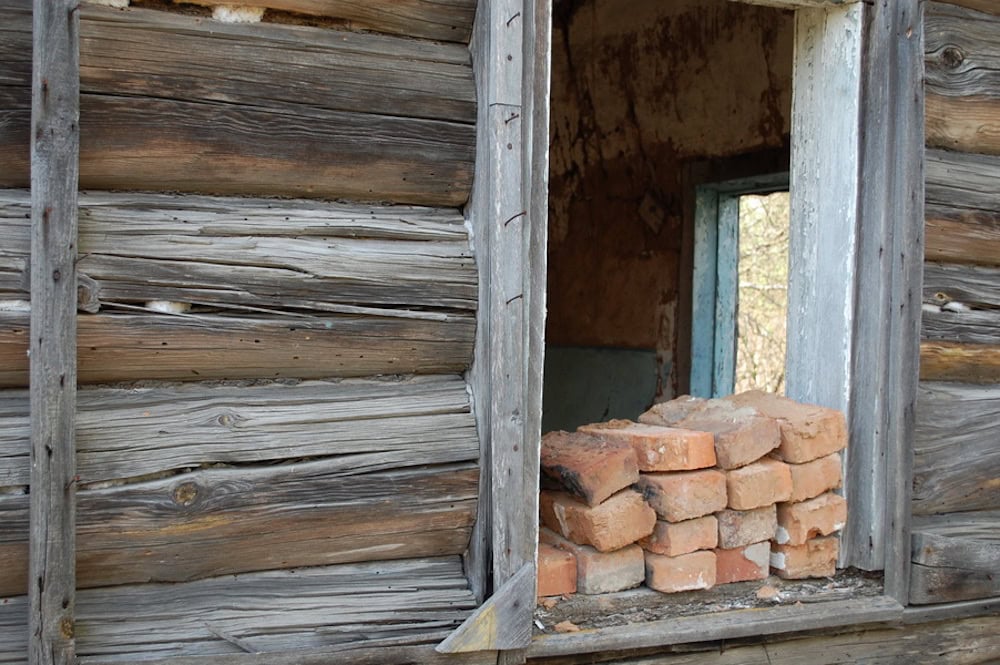
[Image above] Bricks in Belarus territory contaminated by radiation fallout from the 1996 Chernobyl nuclear power plant explosion. Researchers at NC State developed a technique that can characterize radioactive sources in brick even when the original radioactive source is no longer there. Credit: Ilya Kuzniatsou, Flickr (CC BY 2.0)
The scientific method is a lot like bricklaying—individual pieces are laid down one by one until you’ve created a solid wall of evidence.
Though this comparison is simile to most researchers, the process is quite literal for researchers at North Carolina State University. They built on research from the past few years to create a literal wall of bricks—but not for the reasons you may think.
This year marks the 50th anniversary of the Treaty on the Nonproliferation of Nuclear Weapons, a landmark international treaty to prevent the spread of nuclear weapons and weapons technology and achieve worldwide nuclear disarmament. Since the Treaty entered into force in 1970, a review of the Treaty’s operations takes place every five years (though this year’s conference is postponed due to the coronavirus pandemic).
There are many facets to achieving nuclear nonproliferation, from nations without nuclear weapons committing not to acquire them to nations with weapons committing to eliminate them. And to verify such commitments are upheld, nuclear nonproliferation authorities must be able to detect and characterize radioactive sources, including a source’s size, location, and type.
However, modern techniques for nuclear material detection require some combination of real-time measurement and/or sampling of the material. In other words, some or all of the nuclear materials have to be present to be detected. But what if the nuclear material was removed from the area—is there a way to detect its previous presence?
This question is what kicked off the brick research at NC State.
In 2017, NC State associate professor of nuclear engineering Robert Hayes and Oklahoma State University research assistant professor Sergey Sholom published a paper showing retrospective imaging of nuclear materials is theoretically possible.
An NC State press release explains how their technique—which is based on solid state retrospective dosimetry techniques—works.
“The technique takes advantage of the fact that radioactive material changes the arrangement of valence electrons—or outer electrons—in insulator materials, such as brick, porcelain, glass—even hard candy,” the press release says. “By taking samples of multiple materials in a room, applying conventional radiation dosimetry techniques, and evaluating how the electrons at those defect sites are organized, researchers can determine the presence and strength of any nuclear materials that were in that room.”
In 2018, Hayes and NC State graduate research assistant Ryan O’Mara validated the technique using bricks. In particular, they showed their technique greatly simplified the sample preparation process.
“For [retrospective dosimetry luminescence methods] it is, in general, beneficial to have samples that are optically transparent and very small in size such as granular material. As a result, the sample preparation generally includes crushing the samples and treating them with harsh chemicals,” they write in the paper. They demonstrated, however, that “acceptable dose deposition profiles can be obtained from minimally prepared brick samples of small grain sizes.”
Although the technique succeeded in characterizing the energy distribution of a radiation source in brick, Hayes said in the press release that this validation was not the final step. “…we are already working to demonstrate that we can use the technique as a three-dimensional ‘gamma camera,’ giving us the ability to capture the dimensions of the source or sources,” he said.
And that brings us to their recent study, published in Radiation Measurements this month.
“Our new work effectively shows that we could take an array of bricks and turn them into a gamma ray camera, characterizing the location and distribution of a radiation source,” Hayes says in the new press release.
For this study, Hayes and O’Mara did not use actual bricks—they instead used commercial dosimeters. But they say the results still strongly suggest they could do the same with bricks.
“That’s because the silicates in brick—such as quartz, feldspars, zircons, and so on—are all individual dosimeters. It is a tedious process to remove those grains from the brick for measurements, but we have done it multiple times,” Hayes says in the press release. “For the goals of this new research, it wasn’t necessary to use brick—we’ve already shown we can do that. This was simply a question of determining how much information we could glean from this approach.”
They found quite a bit of information can be gleaned—they not only accurately predicted the location of weapons-grade plutonium but even the radius of the source.
There still is work to be done to advance this technique, such as determining proper protocols for sample acquisition, transport, and storage and accounting for errors/bias in the models used to characterize radiation. Regardless, “This ability for three-dimensional imaging is a novel capability,” O’Mara says in the press release.
The 2017 paper, published in Health Physics, is “Retrospective imaging and characterization of nuclear material” (DOI: 10.1097/HP.0000000000000680).
The 2018 paper, published in Health Physics, is “Dose deposition profiles in untreated brick material” (DOI: 10.1097/HP.0000000000000843).
The 2020 paper, published in Radiation Measurements, is “Retrospective characterization of special nuclear material in time and space” (DOI: 10.1016/j.radmeas.2020.106301).