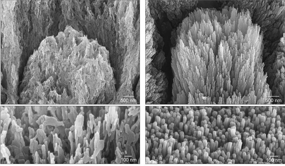
[Image above] Electron microscopy images of a tooth with demineralized enamel showing eroded apatite crystals (left) and a similar demineralized tooth after a 2-week treatment showing epitaxially regenerated enamel crystals (right). Credit: Hasan et al.(opens in new tab), University of Nottingham
Although dentistry dates to 7000 BCE(opens in new tab), researchers continue to develop materials and technologies to improve our standard of dental care. One persistent problem that finally may be solved is that of irreversible enamel loss(opens in new tab), which is one of the main oral diseases(opens in new tab) contributing to the estimated $710 billion(opens in new tab) per year spent on oral treatment and productivity losses.
Natural dental enamel consists of several layers of carbonated hydroxyapatite nanocrystals around 50 nanometers in diameter. This structure results in mechanical properties, such as hardness and wear resistance, that allow enamel to withstand long-term chewing and exposure to the harsh oral environment. Tooth enamel also contains amelogenin, a special type of protein that makes up about 90% of the enamel’s matrix and provides support to surrounding tissues.
Currently, there is no product on the market that can be applied on demineralized or eroded enamel that produces the same structure and properties of natural enamel. Methods based on organic–inorganic nanocomposites, including amelogenin analogues, peptides, and calcium-phosphate particles, have been able to mimic natural enamel to a certain extent. However, their fabrication conditions are not applicable to clinical settings because they generally rely on toxic components, long application times, and/or offer limited control of the mineralization process.
Researchers have spent decades trying to develop a solution without these drawbacks. One research team led by the University of Nottingham in the U.K. may finally have developed a solution, as described in a November 2025 open-access paper(opens in new tab) published in Nature Communications.
The road to this breakthrough was a long one filled with a lot of collaboration, explains senior author Alvaro Mata(opens in new tab), chair of the Department of Biomedical Engineering & Biomaterials at the University of Nottingham, in an email.
“We have been working on tissue regeneration for more than 17 years and began focusing on dental tissues around 2015,” he says. “Much of our initial work was inspired by pioneering scientists around the world, some of whom we have had the chance to collaborate with. For decades, they have been helping us to understand how natural enamel forms, how enamel proteins work, how molecules can be assembled into functional ensembles, how to guide organic–inorganic interactions, how biomineralizing processes can be regulated, etc.”
With a better understanding of these phenomena, the researchers turned to genetically engineered protein polymers used in tissue engineering called elastin-like recombinamers (ELRs). The synthesis of ELRs allows exact control over molecular weight, chain architecture, and sequence design, ensuring monodisperse and uniform polymer properties.
The researchers’ ELR consisted of several types of pentapeptides found in elastin-like polypeptides(opens in new tab) and statherin(opens in new tab), an acidic phosphoprotein found in human saliva. These ELRs were engineered into a clinically friendly supramolecular matrix, which incorporated Ca2+ ions and drying to promote the assembly of the ELR molecules into fibrillar structures.
According to the researchers, “the ELR fibrils exhibited similar structural and functional features to amelogenin fibrils, which are critical to develop our natural enamel during infancy. These fibrils contained embed Ca2+ ions which served as nucleation points to promote mineralization, as confirmed by X-ray photoelectron spectroscopy, transmission electron microscopy, scanning electron microscopy, and computer simulations.”
Upon application of the ELR matrix to enamel tissue, epitaxial and organized growth of apatite nanocrystals occurred from the enamel–ELR interface through the ELR matrix, forming an integrated and organized 10-micron thick mineral layer. This layer consisted of approximately 50-nm diameter nanocrystals that were several microns in length and exhibited the typical hexagonal apatite morphology of native enamel.
The team demonstrated that the epitaxial mineralized layer could be grown from dental enamel, independently of the level of enamel erosion. The matrix was highly stable and able to regrow all the anatomical features of natural enamel with high precision and reproducibility, including prismatic, interprismatic, and aprismatic regions. The chemical composition was also similar to native enamel.
Mechanical properties required to survive toothbrushing, chewing and grinding, and exposure to acidic solutions were restored as well. In fact, fatigue resistance was better than native enamel, as demonstrated by an experiment that simulated regular toothbrushing for one year..
The remineralized enamel showed no visual microstructure loss and displayed similar stiffness and hardness after the experiment, compared to those of native enamel. These results were attributed to the more densely packed apatite nanocrystals in the remineralized enamel. Additionally, enhanced chemical stability was attributed to small amounts of fluorine (levels that are normally present in natural saliva).
Besides the impressive mechanical properties, the researchers were able to apply the ELR coating in a uniform manner over large areas and complex tooth structures in just three to four minutes. This ability is another key factor in achieving successful clinical application.
The researchers note that treatment time depends on the specific condition being treated, the oral environment of each person (e.g., quality of the saliva), and other factors. Work continues to optimize the technology so that it can be used to treat different kinds of oral conditions and pathologies.
One of the key advantages of the technology is the capacity to grow an enamel-like layer from exposed dentin. This ability is critical for bonding applications, which include repairing chipped or cracked teeth; filling in gaps between teeth; covering exposed roots; and adhering veneers, crowns, and onlays. The technology could “potentially provide a one-pot solution for the regeneration of dental enamel independently of the level of tooth erosion,” the researchers write.
The researchers launched a start-up company called Mintech-Bio(opens in new tab) to commercialize the technology, with the first product expected to be on the market in 2027. A clinical trial is also underway in the U.K.
“For all [our products], our main goal is to make them accessible, practical, affordable, and clinically friendly,” says Mata in the email.
The open-access paper, published in Nature Communications, is “Biomimetic supramolecular protein matrix restores structure and properties of human dental enamel(opens in new tab)” (DOI: 10.1038/s41467-025-64982-y).